Branch-specific plasticity explains distal enrichment of retinotopically displaced inputs in visual cortex
bioRxiv · 2026
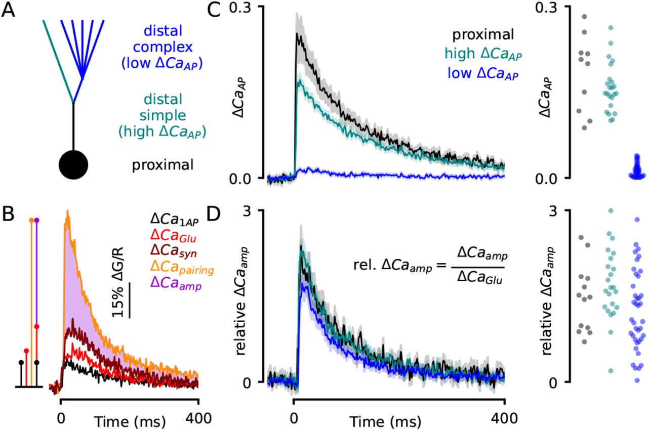
In our earlier eLife study, we found that a spike traveling back into a neuron’s dendrites triggers much less calcium entry in some branches than others, even at a similar distance from the cell body. Figure 1 shows the observation that motivated this preprint. In dendrites with more complex branching, a spike on its own evoked little calcium entry, but it still boosted the calcium signal through NMDARs when paired with synaptic input. That difference led us to model synapses on these branches as less prone to weakening while still able to strengthen, due to the fact that VGCC dependent influx leads to depression, whereas NMDAR dependent influx leads to potentiation.
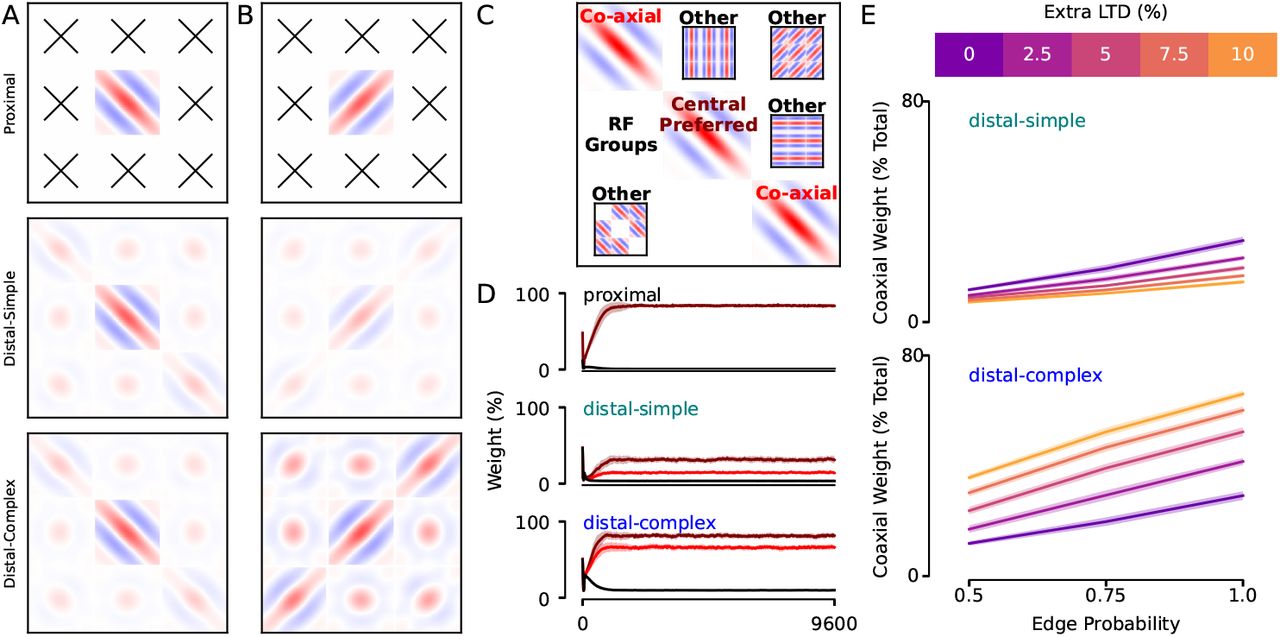
We modeled visual input and plasticity and replicated a finding observed in experimental measurements of mouse visual cortex. When synapses on complex distal branches were less prone to weakening, they retained inputs that preferred the same orientation as the neuron but responded to different points along the same edge. This provides a specific explanation for why inputs are more likely to be retinotopically displaced distally, and provides the additional hypothesis that this phenomenon should be enriched on complex branches.